Our Services  Pre-Natal Diagnosis
Routine Fetal Morphology Scan (18-22 weeks of gestation)
Pre-Natal Diagnosis
Routine Fetal Morphology Scan (18-22 weeks of gestation)
A fetal morphology scan aims to carry out a thorough check of the structure of the fetus. Most babies are normal and the parents can be reassured with the normal ultrasound findings. In case a baby has some structural defects, it might be important to find out before he or she is born. By knowing the abnormalities, prenatal diagnosis might be possible and the parents, the obstetrician and the paediatricians might be better prepared for the birth of such affected child. For certain major congenital defects, the parents might have the option of a termination of the pregnancy if prenatal diagnosis can be established.
What are the limitations of morphology scan?
Not all congenital defects can be picked up on the routine morphology scan. It can pick up around 70-80% major congenital defects. The pick-up rate depends on a number of factors, including the severity and nature of the abnormality, whether the fetal position is optimal for scanning, women’s factors such as whether the tummy is thick or there is surgical scar, the quality of the ultrasound machine and whether the medical personnel are experienced in or have received adequate training in this practice.
Is a morphology scan safe to the fetus?
Diagnostic ultrasound has been used since 60’s. Based on numerous follow-up studies on children who were scanned before birth, there is no evidence that in-utero exposure of antenatal ultrasound is associated with any undesirable biological effects.
Ultrasound Images
First Trimester
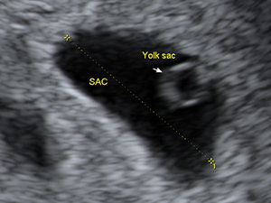 Gestational sac and yolk sac at 6 weeks
 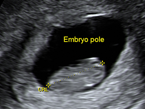 The crown-rump length of the embryo at 9 weeks
 The crown-rump length of the fetus at 12 weeks
 The nuchal translucency thickness in the first trimester
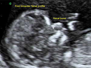 Facial profile in the first trimester
 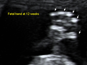 Fetal hand at 12 weeks
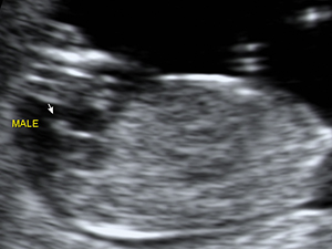 Male genital organ (arrow) at 12 weeks
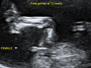 Female genital organ (arrow) at 12 weeks
 Second Trimester - Head and Face
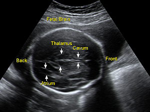 Fetal brain in the second trimester
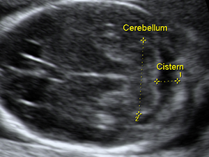 Fetal cerebellum at 17 weeks
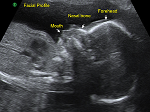 Facial profile in the second trimester
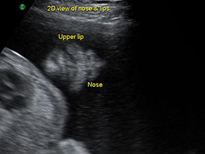 Fetal nose and lips in the second trimester
 Spine
 Fetal spine in the second trimester
 Abdomen
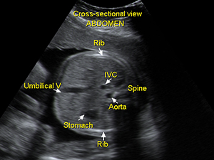 Cross-sectional view of the fetal abdomen
 Umbilical cord insertion of the fetal abdomen
 Fetal bladder
 Two fetal kidneys
 Heart
 The 4-chamber view of fetal heart
 The aortic outflow tract from the fetal heart
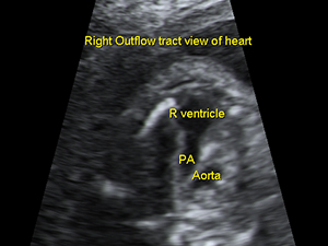 The pulmonary outflow tract from the fetal heart
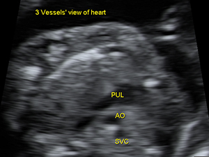 The 3 vessels’view of the fetal heart
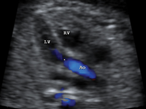 Aortic outflow tract with colour indicating the flow
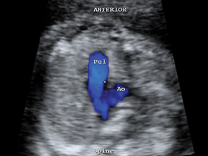 Colour flow of both pulmonary artery and aorta
 Limps
 The fetal hand at 20 weeks
 Gender - Male
 Male genital organ (arrow) at 18 weeks
Male genital organ (arrow) at 18 weeks Gender - Female
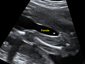 Female genital organ (arrow) at 20 weeks
Female genital organ (arrow) at 20 weeks