


You are here
Introducing Definition Flash CT Scanner - Flash Speed • Lowest Dose • Dual Energy
2010-01-26
Somatom Definition Flash
There are three unique features to the scanner: Lowest Dose, Flash Speed and Dual Energy scanning.
1. Lowest Dose is achieved through:
a) Very fast speed
b) Adaptive dose shield
c) X-care (organ sensitive dose protection)
d) Dual energy scanning
e) Iterative reconstruction
When scanning at very fast speed, X-ray is minimized.
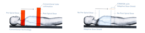
Adaptive dose shield automatically places shields to block out unnecessary X-ray at the top and bottom of the scan (fig 1).
Fig 1.
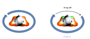
X-care switches off the X-ray in organ sensitive areas e.g. the eyes, thyroid glands and the breasts (fig 2).
Fig 2.
Dual energy scanning may eliminate the non-contrast scan, cutting 30% of the dose.
Iterative reconstruction enables noise reduction in the image, again cutting 30 to 50% of dose.
For example, cardiac scans are performed with ¼ of a heart beat resulting in less than 1 mSv dose (fig 3). This ultra low dose is now routine for patients with heart rates less than 65 bpm. For those with higher heart rates, the dose is still less than that of conventional coronary angiography (which is approximately 5 to 9 mSv).
Fig 3.

2. Flash Speed:
The World Fastest CT scanner with true temporal resolution of 75 ms. Sub-second cardiac scans are performed. Entire thorax scanned in 0.6 seconds with or without breath-holding eliminating motion artefacts (fig 4). Triple rule out (coronary arteries, pulmonary arteries and aortogram) in less than 5 seconds. Whole body scan of 5 to 6 seconds. This scanner is therefore ideal for scanning children, eliminating the need for sedation (fig 5). It is also ideal for intubated, critically ill, or trauma patients.
Fig 4.
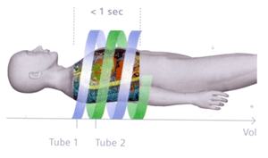
Fig 5.

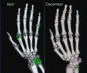
3. Dual Energy scanning:
Dual Energy scanning represents a quantum leap in CT technology. Single energy scans provide only morphological information. Scanning with 2 X-ray tubes emitting two different energies (80 KV and 140 KV) enable functional imaging. One can characterize and visualize the chemical composition of different materials (figs 6 & 7). This leads to unsurpassed diagnostic accuracy as well as opens the door to many new as yet undetermined clinical applications for decades to come.
Fig 6.
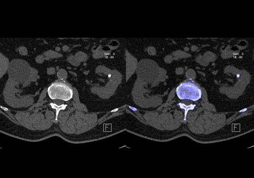
Fig 7.
There are three unique features to the scanner: Lowest Dose, Flash Speed and Dual Energy scanning.
1. Lowest Dose is achieved through:
a) Very fast speed
b) Adaptive dose shield
c) X-care (organ sensitive dose protection)
d) Dual energy scanning
e) Iterative reconstruction
When scanning at very fast speed, X-ray is minimized.
Adaptive dose shield automatically places shields to block out unnecessary X-ray at the top and bottom of the scan (fig 1).
Fig 1.
X-care switches off the X-ray in organ sensitive areas e.g. the eyes, thyroid glands and the breasts (fig 2).
Fig 2.
Dual energy scanning may eliminate the non-contrast scan, cutting 30% of the dose.
Iterative reconstruction enables noise reduction in the image, again cutting 30 to 50% of dose.
For example, cardiac scans are performed with ¼ of a heart beat resulting in less than 1 mSv dose (fig 3). This ultra low dose is now routine for patients with heart rates less than 65 bpm. For those with higher heart rates, the dose is still less than that of conventional coronary angiography (which is approximately 5 to 9 mSv).
Fig 3.
2. Flash Speed:
The World Fastest CT scanner with true temporal resolution of 75 ms. Sub-second cardiac scans are performed. Entire thorax scanned in 0.6 seconds with or without breath-holding eliminating motion artefacts (fig 4). Triple rule out (coronary arteries, pulmonary arteries and aortogram) in less than 5 seconds. Whole body scan of 5 to 6 seconds. This scanner is therefore ideal for scanning children, eliminating the need for sedation (fig 5). It is also ideal for intubated, critically ill, or trauma patients.
Fig 4.
Fig 5.
3. Dual Energy scanning:
Dual Energy scanning represents a quantum leap in CT technology. Single energy scans provide only morphological information. Scanning with 2 X-ray tubes emitting two different energies (80 KV and 140 KV) enable functional imaging. One can characterize and visualize the chemical composition of different materials (figs 6 & 7). This leads to unsurpassed diagnostic accuracy as well as opens the door to many new as yet undetermined clinical applications for decades to come.
Fig 6.
Fig 7.


