Department of Nuclear Medicine & Positron Emission Tomography ❯ Our Research & Resources
Our Research
In the recent decade, the Hospital has put more emphases and resources on applied clinical research and its contribution to the health services of patients in Hong Kong. Medical imaging with Positron Emission Tomography/Computed Tomography (PET/CT) is found valuable in oncology, cardiology and neurology. Our department is internationally known for the following research fields: hepatocellular carcinoma (HCC) and other liver pathology including cholangiocarcinoma, focal nodular hyperplasia (FNH), hemangioma, adenoma, metastatic tumors; renal cell carcinoma, angiomyolipoma; myeloma; Alzheimer and neurodegenerative diseases, etc. All of our research is most notable to correlate with clinical diagnosis capability. We have published many first class papers in top medical imaging journals (including one that was rewarded an invited commentary and one cover article) and provided keynote lectures all over the world. Over 100 international publication were achieved in the past 10 years. Our department also won the International Best Abstract Awards from the Society of Nuclear Medicine and Molecular Imaging in 2015, 2016, 2018 and 2022, 4th time out of the past 8 years since the establishment of the award.
HCC is one of the most common malignancies worldwide. Its occurrence is much higher in the Asian and African. Accurate differentiation and staging of HCC may increase the chance of curative treatment, affect the patient management, lower the rate of recurrence and, hopefully, prolong survival. Our center is the first institute in the world using dual-tracer (C-11 Acetate and F-18 FDG) to detect HCC, improving the detection sensitivity from 40-50% to nearly 100%. Our center is the only center in Hong Kong using dual-tracer PET (C-11 PIB and F-18 FDG/ F-18 TAU) and customized region-based quantitative analysis for the diagnosis of Alzheimer's disease, which could detect dementia at an early stage and improve the diagnostic sensitivity and specificity compared with 18F-FDG-alone PET.
“There is a recent important publication and decision made by the NIA-AA (National Institute on Aging and Alzheimer's Association) jointly by the world’s leading neurologists including Mayo, Harvard, Stanford, Washington U, U Penn, Chicago, UC Berkeley, UCSF, FDA, as well as those from Germany, Barcelona and Australia, on the revised definition on Alzheimer's disease (AD).
The NIA-AA Research Framework has arrived a new but practical definition for AD, which was previously a syndrome diagnosed by a set of clinical criteria, it is now defined by “Biomarkers”. These biomarkers are categorized into 3 groups: (1) beta-Amyloid, (2) pathologic Tau, and (3) neurodegeneration. In short, they are called “ATN”. Both Amyloid (A) and Tau (T) are the abnormal protein deposits that define AD as a unique neurodegenerative disease that can lead to dementia.
As PET imaging with PIB and T-807 are the surrogate biomarkers for Amyloid and Tau proteins, respectively, the NIA-AA’s decision to shift the definition of AD to a biological construct is a very logical and important move for the future of PET in neurological imaging.
This paper serves to guide the future direction of neurologic PET as well as to establish the recognition of molecular PET in AD, which is one of the most important diseases of human aging in the coming years.”
The quality of our team and the fruitfulness of our research outputs make our service distinguishing and unique in Hong Kong.
Dr. C.L. Ho
Head, Department of Nuclear Medicine & PET
HKSH Medical Group
Research Highlights
Hepatocellular carcinoma
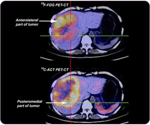
In the same HCC, C-11 ACT and F-18 FDG can be taken up by different parts of the same tumor due to different cellular differentiation
Neuroendocrine tumour

F-18 DOPA scan demonstrates multiple neuroendocrine tumors
Dementia
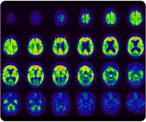
C-11 PIB PET detects typical Alzheimer's disease with amyloid plaque at post cingulate gyrus, frontal lobe, precuneus, parietal lobe and temporal lobe.
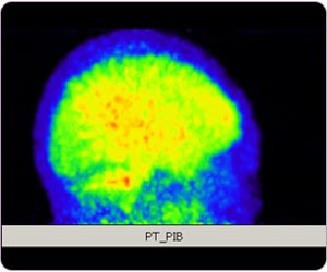
C-11 PIB PET MIP: Alzheimer disease
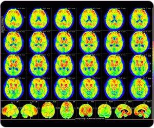
Region-based quantitative analysis for C-11 PIB brain study
Theranostics
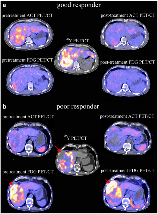
C-11 ACT, F-18 FDG PET/CT and Y-90 microsphere radionuclide therapy
In the same HCC, C-11 ACT and F-18 FDG can be taken up by different parts of the same tumor due to different cellular differentiation
Neuroendocrine tumour
F-18 DOPA scan demonstrates multiple neuroendocrine tumors
Dementia
C-11 PIB PET detects typical Alzheimer's disease with amyloid plaque at post cingulate gyrus, frontal lobe, precuneus, parietal lobe and temporal lobe.
C-11 PIB PET MIP: Alzheimer disease
Region-based quantitative analysis for C-11 PIB brain study
Theranostics
C-11 ACT, F-18 FDG PET/CT and Y-90 microsphere radionuclide therapy
Academic Activities & Output:
| Year | Journal paper | Abstract |
| 2003 | 3 | 5 |
| 2004 | 3 | 1 |
| 2005 | 1 | 5 |
| 2006 | 3 | 2 |
| 2007 | 4 (1 cover article) | 1 |
| 2008 | 1 | 2 |
| 2009 | 4 | 1 |
| 2010 | 4 | 5 |
| 2011 | 4 | 8 |
| 2012 | 3 | 8 |
| 2013 | 2 | 6 (Distinguished Abstract Award) |
| 2014 | 4 | 11 (Best Abstracts Award) |
| 2015 | 3 | 7 (Best Abstracts Award) |
| 2016 | 2 | 4 (Best Abstracts Award) |
| 2017 | 0 | 9 |
| 2018 | 3 | 6 (Best Abstracts Award) |
| 2019 | 4 | 6 |
| 2020 | 2 | 0 |
| 2021 | 4 | 0 |
| 2022 | 3 | 4(Best Abstracts Award) |
Research Highlights (Majority of published full papers are in top 3 medical imaging journals):
- 11C-acetate & 18F-FDG PET/CT for the early diagnosis of HCC, HCC management and prediction of survival (No. of publications: 53)
- 11C-PIB and 18F-T807 PET/CT for the early diagnosis of Alzheimer disease, predicting MCI to Alzheimer disease (No. of publications: 15)
- 11C-acetate & 18F-FDG PET/CT in diagnosis and management of multiple myeloma (No. of publications: 13)
- 11C-acetate & 18F-FDG PET/CT in renal cell carcinoma and predicting survival (No. of publications:6)
Resources
What is Radiation?
Radiation is a way of transmitting energy through high-energy particles or waves. Radiation can be divided into two main categories: electromagnetic radiation and particle radiation.
Electromagnetic radiation refers to energy waves generated by an electromagnetic field, including visible light, radio waves, infrared, ultraviolet, X-rays, and gamma rays. The energy of electromagnetic radiation can range from less than 1 electron volt (eV) to several hundred million electron volts (MeV), and different energy levels of electromagnetic radiation have different characteristics and applications. For example, visible light can be used for illumination, infrared can be used for thermal imaging, X-rays can be used for medical imaging and industrial inspection, and gamma rays can be used for sterilization, radiation therapy, nuclear medicine and positron emission tomography imaging.
Particle radiation refers to high-energy particles released by atomic nuclei or other particles, such as alpha particles, beta particles, and neutrons. These particles have different energies and penetrating abilities. They can cause chemical reactions or radiation damage in materials, and apply in radionuclide therapy for cancer treatment.
Radiation has a wide range of applications in fields such as medicine, industry, and energy, but it can also affect human and the environment if applied inappropriately. Therefore, the safety and monitoring when utilizing radiation is important. In medical applications, it is necessary to fully consider the necessity and weigh the pros and cons, and ensure that the radiation dose does not exceed the safety limit, and measures are taken to protect patients and professional personnel.
Radiation in Daily Life
Radiation is closely related to us in the natural environment, as both air and the earth itself are radioactive. Background radiation is the natural radiation that continuously exists in the environment. The average annual background radiation on Earth is about 1.8-3.6 mSv (millisieverts). The main sources of radiation in the human body are food, water, and air. The radiation dose from a typical isotope myocardial perfusion scan is approximately 10 mSv, the associated risk factor induced by radiation is equivalent to living in air-polluted New York or Boston for about 6 months, serving as a flight attendant in the cabin for about three years, driving motorcycle for 1000km or smoking seven packs of cigarettes. The misuse of radiation can certainly have adverse effects or harm on the human body, but when used under the monitoring of professionals, radiation can effectively improve the quality of human life and contribute to medical, research, and industrial development.
Radiological examinations, treatments, or procedures used for medical purposes must be performed only when the benefits outweigh the risks and are confirmed by a doctor as necessary. Doctors consider the urgency of the patient's need for examination and clinical data, striving to select examinations or treatments with the least risk that can improve or help diagnosis of the patient. Although undergoing radiological scans or treatments may still increase the body's radiation exposure slightly, not following the doctor's advice for examination may lead to misdiagnosis or delayed treatment, resulting in more serious consequences.
Fear of Radiation
Nuclear technology is gradually being widely used in many fields such as medical imaging, radiation therapy, and energy production. Governments around the world have established radiation management agencies and developed radiation safety guidelines, regulations, and policies to monitor and control radiation risks. However, some organizations tend to be overly cautious in their radiation safety and protection policies, even concluding that radiation is harmful and has no benefits, leading to various misunderstandings about radiation among the public and causing a great deal of fear of nuclear radiation.
The Atomic Bomb Survivors Life-Span Study (LSS) is the most comprehensive longitudinal study ever conducted. The LSS data described by an S-shaped dependence on radiation exposure with a threshold of about 200mSv showed that “Linear no threshold hypothesis”-predicted low-dose carcinogenicity is invalid up to approximately 200mSv. Indeed, the curvature showing negative excess relative risk suggested a “hormesis” concept below 200mSv.
Some studies, on the other hand, have suggested that extremely high-dose radiation can cause genetic mutations, thereby increasing the risk of cancer. However, genetic mutations do not necessarily lead to cancer. Genetic mutations are common in the human body because the structure of chromosomes is not stable. Not only can most chromosomes repair themselves, but the number of spontaneous genetic mutations is far greater than those caused by low-dose radiation. The human body has self-adjusting and repairing functions, which are innate disease prevention and resistance abilities and an important mechanism for preventing cancer. Moreover, many recent studies have also shown that low-dose radiation employed in diagnostic imaging range not only does not increase the rate of genetic mutations but also reduces it and can enhance the self-repair function of chromosomes, thereby reducing the risk of cancer.
A Bit More Knowledge, From the Other Side of the World
The worst nuclear accident in history was probably the Chernobyl nuclear power plant explosion in Ukraine in 1986. This deepened people's alertness on radiation and greatly increased their fear of it. The Fukushima nuclear accident in Japan caused by natural disasters in 2011 once again drew public attention to radiation and its effects. Although past studies have shown that radiation can increase the rate of genetic mutations or the risk of cancer, these are all based on the use of extremely high-dose radiation. In fact, low-dose radiation may not necessarily have a negative impact or harm on the human body.
Background radiation is the most common type of radiation that people come into contact with in daily life. The amount of radiation the body receives often depends on different locations, environments, and habits. According to data from the Radiation Health Division, Department of Health, Hong Kong, the average monthly background radiation in Hong Kong is about 150-300 μSv (microsieverts) (about 1.8-3.6 mSv (millisieverts) per year). The average annual background radiation in Yangjiang City, Guangdong Province, China, is about 6.4 mSv (millisieverts), which is about twice or more than that in Hong Kong. After a study involving more than 125,000 people as a control group for 20 years, the results showed that the number of Yangjiang citizens who died of cancer was actually lower than that of the general control group.
In Ramsar (a city in Iraq), the annual background radiation level is one of the highest in the world due to nearby hotsprings and building materials with dissolved radium originating from them. It can reach up to about 100-260 mSv (millisieverts), which is tens to a hundred of times higher than in Hong Kong. This level of radiation exposure is not even reached by a radiation therapist or flight attendant after 38 years of work. However, the high background radiation environment in Ramsar has not had a negative impact on its residents.
In 1983, the radiation steel incident in Taiwan resulted in contaminated buildings, including schools, residences, and office buildings. An estimated 10,000 people were affected, with 1,000 of them accumulating radiation levels of up to 4,000 mSv within 20 years. According to the established international guidelines on radiation protection, the predicted number of cancer death would be 302, with 232 natural cause plus 70 induced by radiation. However, only 7 people actually died of cancer 20 years later, which is only 3% of general cancer death rate.
The negative impact and risks of radiation are deeply ingrained in the public's memory, but most people's fear is not due to the existence of radiation, but rather from the unknown or lack of understanding of the issue, combined with their own imagination, which amplifies their concerns infinitely. In fact, the application of radiation is becoming more and more widespread, and in addition to the negative impact of radiation, understanding more about radiation protection and its benefits may help people to overcome their prejudice against the safe use of radiation and gradually regain confidence.
To those who suffer from radiophobia:
The negative effects of radioactivity have made more and more people worried about the harm caused by radiation, and some even believe that any dose of radiation can cause cancer, and that there is no such thing as a safe radiation level in the world. Indeed, among daily activities, travelling by airplanes or living in mountainous areas increases absorption of cosmic radiation due to high altitude. Consumption of fruits and vegetables will lead to ingestion of natural radioactive isotopes from soil and rocks. Radon gas, which is one of the possible causes of lung cancer, is produced from decomposition of natural radioactive substances and can be acquired through breathing. Moreover, human or animal body itself carries radioactivity and getting close to each other may become a concern for people with radiophobia.