Department of Nuclear Medicine & Positron Emission Tomography ❯ Our Services ❯ Positron Emission Tomography/Computed Tomography (PET/CT)
Positron Emission Tomography/Computed Tomography (PET/CT)


High Sensitivity
Positron Emission Tomography/Computed Tomography (PET/CT) is one of the most effective methods for cancer imaging and diagnosis. During the course of cancer development, metabolic changes always come earlier than structural or morphological changes in tissues and organs. Therefore PET/CT being a powerful camera capable of imaging our metabolic functions, it can facilitate early cancer detection and staging through the identification of the tumor biological characteristic.
High Specificity
PET tracers work as the biological imaging probe. Some of the commonly applied PET tracers include: PET radionuclide labelled deoxyglucose, acetate, DOTA-conjugated peptides, prostatic specific membrane antigen, etc. After injection, the tracer is absorbed based on organs’ structure as well as cell metabolism. The patient is then scanned with the PET/CT scanner to pick up any signals emitted from the tracer and hence the lesion location can be identified and uptake can be quantified. With this unique piece of medical information, we can locate lesions precisely and also help the assessment of treatment effectiveness.
Disease Quantification
PET/CT offers multiple parameters to quantify the cell activity which has a broad clinical application like cancer, cardiac, neurological diseases, etc. Apart from the most common “Standardized Uptake Value (SUV)”, the latest application involves mathematic calculation on the image data to retrieve even more unique dynamic parameters (such as “Patlak Analysis” for absolute metabolic rate, distribution volume of the pharmacology, theranostics and dose planning in radiotherapy). It creates unlimited possibilities in diagnosis and treatment planning.
Revolutionary concept of treatment
Theranostics is a revolutionary concept which brings diagnosis and therapy closer together than ever before. The biological distribution of a specific tracer can be observed from the diagnostic scan and later in the treatment phase, that type of tracer is used as a carrier of a treatment radiation dose targeted accurately to the tumor. As a result, the diagnostic scan serves as a simulation to forecast the possible treatment outcome, i.e., what we see is what we treat. Moreover, the carrier can even deliver the radiation dose to very small size lesion that we cannot see.
Through quantification of absorbed dose from post treatment scan, the treatment effectiveness can be reviewed in a comprehensive way. Examples are Fluorine-18/Lutetium-177 PSMA cancer treatment, Gallium-68/Lutetium-177 DOTA Conjugated peptides and Technetium-99m MAA/Yttrium-90 microspheres liver cancer treatment. Please click here for Radionuclide Therapy.
Through quantification of absorbed dose from post treatment scan, the treatment effectiveness can be reviewed in a comprehensive way. Examples are Fluorine-18/Lutetium-177 PSMA cancer treatment, Gallium-68/Lutetium-177 DOTA Conjugated peptides and Technetium-99m MAA/Yttrium-90 microspheres liver cancer treatment. Please click here for Radionuclide Therapy.
Specialized in Nuclear Medicine
All procedures are supervised by Specialists in Nuclear Medicine, which is different from other local medical institutions. After consultation with patients, our specialist will arrange the most suitable examinations and report findings based on clinical needs.
Multiple PET tracers available in HKSH
Adopting different PET radiopharmaceuticals specified to different types of cancer cells is the unique feature of PET/CT scan. Our team is specialized in synthesizing close to 20 radiopharmaceuticals tagged with F-18, C-11 or Ga-68 for clinical use. Common examples are:
Neuroendocrine Tumor
Dual tracer PET/CT (F-18 FDG and Ga-68 DOTATATE) are used for the metastatic evaluation of neuroendocrine tumor.
Dual tracer PET/CT (F-18 FDG and Ga-68 DOTATATE) are used for the metastatic evaluation of neuroendocrine tumor.
 [F-18] FDG |
 [Ga-68] DOTATATE |
Liver Cancer
Multiple tracers are used to differentiate biochemical properties of liver lesion – Cancer Associated Fibroblast (Ga-68 FAPI uptake)
[C-11] Acetate and [F-18] FDG for hepatocellular carcinoma cellular differentiation (well and poorly differentiated)
Multiple tracers are used to differentiate biochemical properties of liver lesion – Cancer Associated Fibroblast (Ga-68 FAPI uptake)
 [F-18] FDG |
 [C-11] Acetate |
 [Ga-68] DOTATATE |
 [Ga-68] FAPI |
[C-11] Acetate and [F-18] FDG for hepatocellular carcinoma cellular differentiation (well and poorly differentiated)
 [C-11] Acetate |
 [F-18] FDG |
Myeloma
 [C-11] Acetate |
 [F-18] FDG |
Prostate Cancer
 [F-18] PSMA |
Brain Tumors
 [F-18] FDG |
 [C-11] Choline |
Neurology
 [C-11] PIB |
 [C-11] Raclopride |
 [F-18] DOPA |
State-of-the-art technology in HKSH
Our Centre has acquired the first innovative digital SiPM PET/CT scanner in China to provide revolutionary breakthrough in PET imaging accuracy. This innovative technology precisely quantifies cell metabolism according to physiologic characteristics and molecular biochemical properties, which is applied to different clinical specialties such as oncology and neurology. Please click here for details.
Oncology
The followings are enquiries on PET/CT related oncology cases:
PET/CT Simulator for Radiotherapy Treatment Planning


PET/CT Simulation is a one-stop imaging facility for both metabolic evaluation and CT simulation in radiotherapy treatment planning. The efficacy of radiotherapy relies on maximizing radiation dose to tumor and preserving normal tissues. Click here for more details.
- How can we know if one has hidden cancer, and if so, its location?
PET/CT can detect and locate primary tumor and occult malignancy by limited wholebody scanning. - How can we tell if the tumor is benign or malignant?
Malignant tumor cells are approximately two to ten times more active than normal cells. They show a higher metabolic rate with rapid cell division cycle. With the use of appropriate metabolic and/ or receptor tracers to probe the tumor, distinct “hot spots” according to the phase of cell differentiation based on biochemistry and receptor density can be seen. - Has the cancer spread (ie. Staging of cancer)?
Limited wholebody scan includes multiple systems and organs of our body. Any metastatic lesions can be detected easily. - How to find out if someone is suitable to take the radionuclide therapy? What is the preparation?
It is necessary to undergo a PET/CT scan or SPECT/CT scan (using specific tracer) to investigate the nature of cancer cell type. - How can we know if the cancer treatment is effective?
After systemic cancer therapies (chemotherapy, hormonal therapy, targeted therapy & immunotherapy), biochemical and functional changes of lesions always appear earlier than structural changes. By comparing the tracer percentage absorption in lesions in pre and post treatment PET/CT or SPECT/CT scans, the effectiveness of treatment can be assessed at a much earlier stage and hence referral doctors can make further plan for cancer management.
PET/CT Simulator for Radiotherapy Treatment Planning
PET/CT Simulation is a one-stop imaging facility for both metabolic evaluation and CT simulation in radiotherapy treatment planning. The efficacy of radiotherapy relies on maximizing radiation dose to tumor and preserving normal tissues. Click here for more details.
Myocardium Viability Evaluation
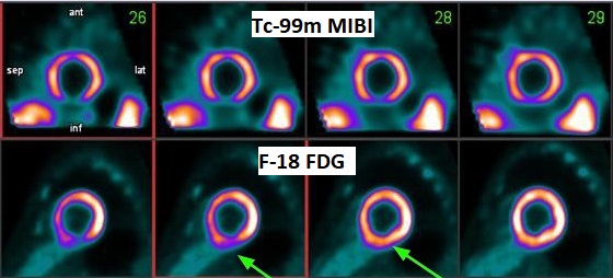
PET/CT and nuclear medicine scans can work together in assessing coronary heart disease. PET/CT scan is particularly helpful in determining whether there is any reversible occlusion (hibernating myocardium). This kind of myocardium still metabolizes glucose even though it seems to be infarcted. With modern advances in cardiology, such hibernating myocardium stands a very good chance of regaining function after angioplasty or cardiac by-pass surgery.
Neurological Diseases
- Dementia

PET/CT scans with special tracers can show 3-dimensional images of blood flow and biomolecules deposition in the brain for diagnosis and monitoring the therapy progress of various neurological diseases, such as Alzheimer's, Dementia, Parkinson's disease, Huntingdon's chorea and autism. In 2018, the National Institute on Aging-Alzheimer's Association (NIA-AA) had revised the definition of Alzheimer's Disease (AD) from a syndrome diagnosed by a set of clinical criteria to a biological construct using abnormal protein deposits to define AD as a unique neurodegenerative disease that can lead to dementia. Our hospital has almost 20 years of experience in producing specific tracer to detect abnormal protein deposition in the brain.
- Epilepsy


For some types of epilepsy patients, PET/CT scans can localize the epilepsy-causing focus in the brain. Modern stereotaxic neurosurgery can then cure epilepsy permanently by excising the focus.
Scan Preparation & Procedures
Is it true that a PET/CT scan will deliver a large amount of radiation to my body?
Generally, the radiation dose of different diagnostic imaging modalities is very safe. Most people understand cancer cells can be destroyed by high dose delivered in radiation therapy over cycles and PET radiotracer contains a radiation dose 10,000 times less than those of radiotherapy. Evidence from “Life span study” (LSS) of Japan atomic bomb survivors from 1958-2009 showed no excess relative risk was found in the survivors compared with the non-exposed control group for all-solid-cancer death below 180mSv. The LSS data showed that the low-dose carcinogenicity as predicted by the Linear No Threshold Hypothesis is invalid up to approximated 200 mSv. Protective cellular biological responses occurred in human bodies with self-repair mechanism, antioxidant defense and immune-system response. Since PET tracers will decay very quickly within few minutes or hours, and appropriate dose of radiation will be used in PET/CT scan based on clinical needs to achieve diagnostic quality images, radiation dose is not a major concern.
How many PET/CT scan can be taken within a year?
There isn’t a distinct limit since the rationale of considering a PET/CT scan mainly based on the clinical need of the patient instead of the radiation dose. It is important not to delay the diagnosis and hence the treatment. If you want more information about radiation, our professional staff will explain to you during your visit.
Is the PET tracer same as the contrast media?
No. Our PET tracers are radioactive biomolecules produced in-house with medical cyclotrons, inside, licensed radiopharmacy laboratories by a team of radiochemist expertise led by a chartered chemist and NM specialist. The production process is strictly monitored under statutory regulations. Since their molecular structure and biological effect are similar to those biomolecules inside our body, they can be absorbed by our cells safely and naturally. Tracer distribution representing cell function can be visualized by PET/CT scanner.
Will PET tracer induce allergy?
The nature and characteristic of PET tracers are very similar to the biological molecules in our body. It is absolutely safe to take a PET/CT scan. Different from iodinated contrast media, physiological elements such as glucose, fatty acid, phospholipid or protein is used. Occurrence of allergic response or discomfort is unlikely. It is unnecessary to carry out contrast allergy assessment beforehand.
Why contrast media is NOT injected during PET/CT scan in our department?
Adopting close to 20 types of PET radiopharmaceuticals specified to different types of cancer cells based on biochemical metabolism or receptor density at molecular level is the unique feature of PET/CT scan in HKSH. PET/CT “fusion image” combines data from the two imaging modalities, with PET providing cellular functional information while CT applies attenuation correction for quantification of PET data on tumour pathophysiology. The presence of iodinated CT contrast media will affect accuracy of PET quantitative data (such as SUV) during interpretation of images. Moreover, kidney function may also be hindered by contrast media. Iodine content in contrast media may induce allergic reaction. Patient will also receive more than double amount of unnecessary radiation dosage, if an extra wholebody contrast CT scan comes along with the plain PET/CT scan.
Except for the 18-Fluorodeoxyglucose (FDG), do I need other PET tracers?
As more than half of the cancer cell entities will absorb glucose for metabolism, 18-Fluorodeoxyglucose (18F-FDG) is most commonly used for oncology assessment. Non-FDG tracers such as acetate, prostate specific membrane antigen (PSMA), DOTA-Conjugated peptides, fibroblast activated protein inhibitor (FAPI), etc. will be utilized in specific cancer types as decided by Nuclear Medicine Specialists based on cancer pathology and staging.
My doctor requested a digital copy of scan images and report, do you have such service?
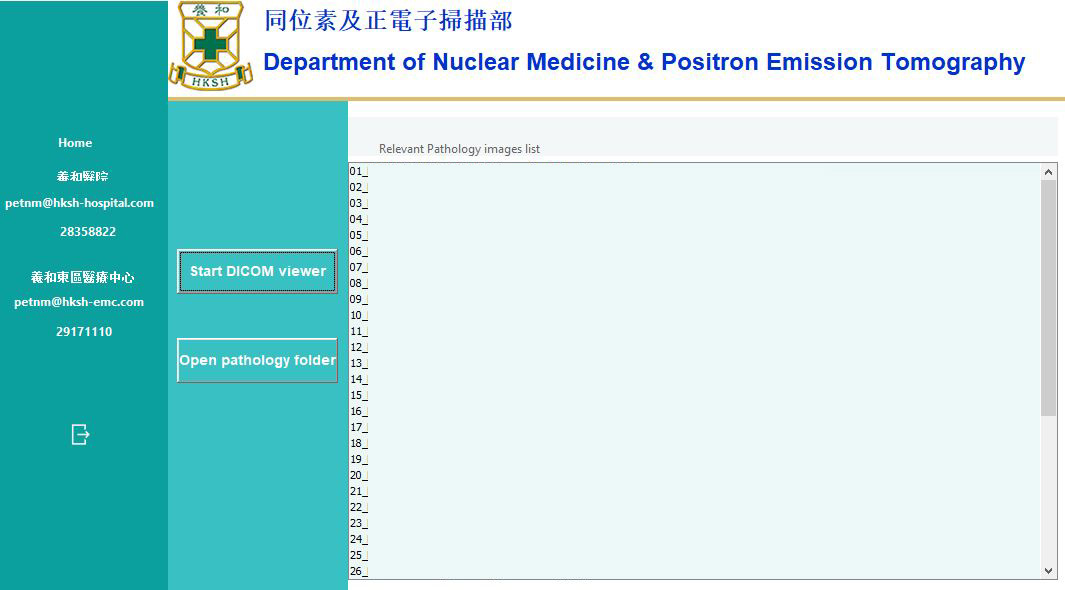
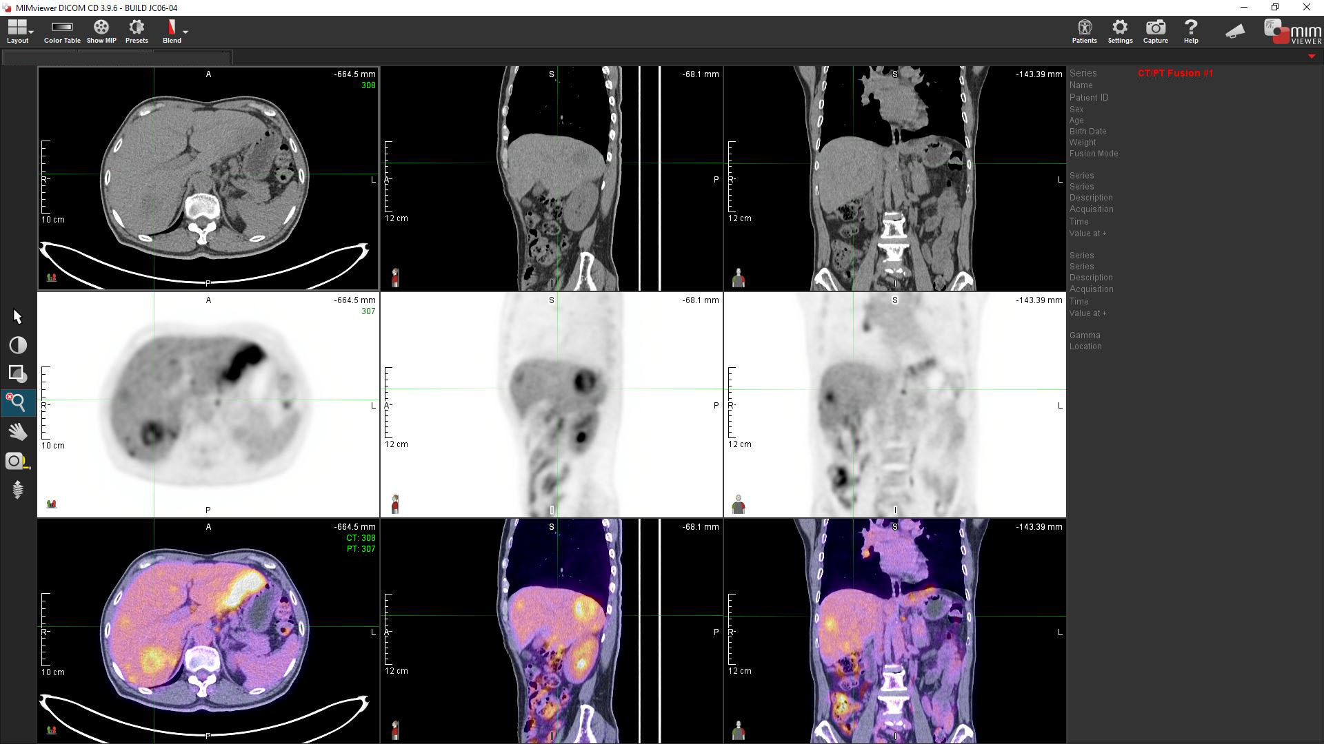
A color-printed booklet with a Write-Once USB drive will be provided as a standard package. All the scan data and relevant pathology images captured by nuclear medicine specialist are included. Using the MIM Viewer software provided, referral doctors or patients can view the images with their own computer. You can also request our staff to upload your images and reports to the Electronic Health Record Sharing System (eHRSS) run by the HKSAR Government to facilitate information sharing among participating hospitals and clinics including the Hospital Authority.

PET/CT ❯

Nuclear Medicine ❯

Radionuclide Therapy ❯

Radiopharmaceutical Dispensing ❯